
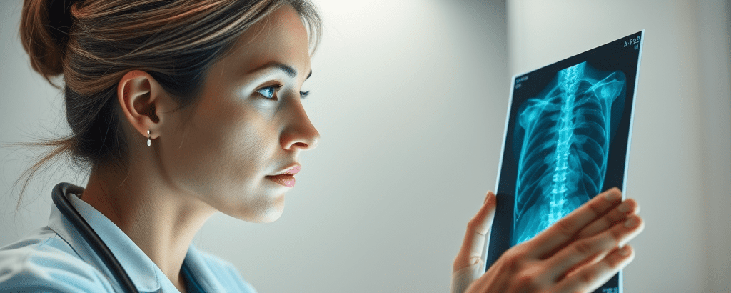
X-rays are an indispensable part of our lives. They are used everywhere. Be it medicine, security or industrial applications. Their journey from an accidental discovery in the laboratory at the University of Wurzburg to its profound impact on humanity is a tale of curiosity, persistence, and innovation.
In this story, I take you through the evolution of X-rays and the legacy of Wilhelm Rontgen, the father of X-rays.
Importance of X-Rays
X-rays allow us to peek inside human bodies and other objects by providing clear images of the internal structures without any invasive procedure. This enables physicians to diagnose bone fractures, ligament injuries, lung infections and other ailments.
X-rays help security professionals detect hidden threats in luggage and personal belongings. They can detect hidden objects, weapons, and contraband. This ensures safety at airports, metro and railway stations and other similar public spaces.
These dual applications highlight the versatility of X-ray technology. Bridging the gap between science and society. A testament to technology addressing real-world challenges effectively.
Wilhelm Rontgen: The Inventor
Wilhelm Conrad Rontgen, the German physicist, was born on March 27, 1845, in Lennep, Prussia. Rontgen was a curious and studious child and had an aptitude for science and engineering. He pursued his education at the Polytechnic School in Zurich, Switzerland, earning a degree in mechanical engineering.
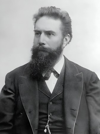
Rontgen’s passion for physics grew during his academic journey, leading to a fruitful career in teaching and research. His expertise and dedication earned him professorships at the University of Wurzburg in Germany, where he would later make his groundbreaking discovery.
The Invention of X-Rays
On November 8, 1895, Rontgen was experimenting with Cathode Rays in his laboratory at the University of Wurzburg. He had been using a glass vacuum tube (Crookes Tube) for this experiment. Suddenly he noticed that a nearby screen coated with barium platinocyanide began to glow. Although no direct light was falling on it. Intrigued, Rontgen began investigating.
Soon he discovered that rays generated in this experiment could pass through substances including human tissue, but not bones and metals. He named these mysterious rays “X-rays”. As always, the ‘X’ denoted the ‘unknown’ in this story. X-rays also produced shadow-like images on photographic plates.
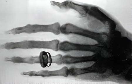
The first X-ray image was a groundbreaking moment. Rontgon captured the bones of his wife Anna’s hand, clearly showing her skeletal structure and her wedding ring. This image, symbolic of the hidden becoming visible, marked the dawn of a new era in medical science. Rontgen’s willingness to share his findings ensured that his discovery was quickly adopted.
Early Use
Doctors quickly realised the potential of X-rays to visualise fractures, tumours, and foreign objects within the body without invasive surgery. The ability to see inside the human body drastically changed medical practices and improved patient outcomes. The early use of the X-rays was to locate bullets in wounded soldiers and diagnose bone fractures.
X-rays also found use in industrial applications. In inspecting the integrity of materials and components in manufacturing. X-rays allowed the detection of defects and irregularities, ensuring higher quality and safety standards.
Despite benefits, early X-rays posed significant health risks. The dangers of ionising radiation were not known then. Prolonged radiation exposure caused burns and other complications among the early users. As time progressed protective measures, such as lead aprons and shields, were introduced to make X-rays safer.
Transforming Medical Diagnosis
X-rays revolutionised medical diagnosis by providing a powerful tool for visualizing internal structures. A few key applications included:
- Fracture Detection: The gold standard for identifying broken bones and joint dislocations.
- Disease Diagnosis: Pneumonia, tuberculosis, kidney stones etc. could be detected with unprecedented accuracy.
- Dental Imaging: X-rays allowed dentists to identify cavities, root infections, and other oral health issues.
The accurate and improved diagnostic aided by the X-rays paved the way for life-saving treatments.
Applications in Security and Travel
The application of X-rays in security began in the mid-20th century, initially in aviation. The ability of X-rays to penetrate solid objects made them ideal for inspecting luggage and cargo without the need for manual searches. They are used for:
- Baggage Screening: X-ray scanners scan luggage and carry-on bags. They detect prohibited items such as weapons, explosives, and contraband.
- Body Scanners: Body scanners provide detailed images of passengers, revealing concealed weapons and harmful objects. This ensures that no dangerous items are carried onto planes.
- Cargo Inspection: Cargo containers are scanned to identify illicit substances and ensure the safety of goods transported across borders.
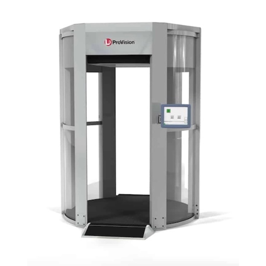
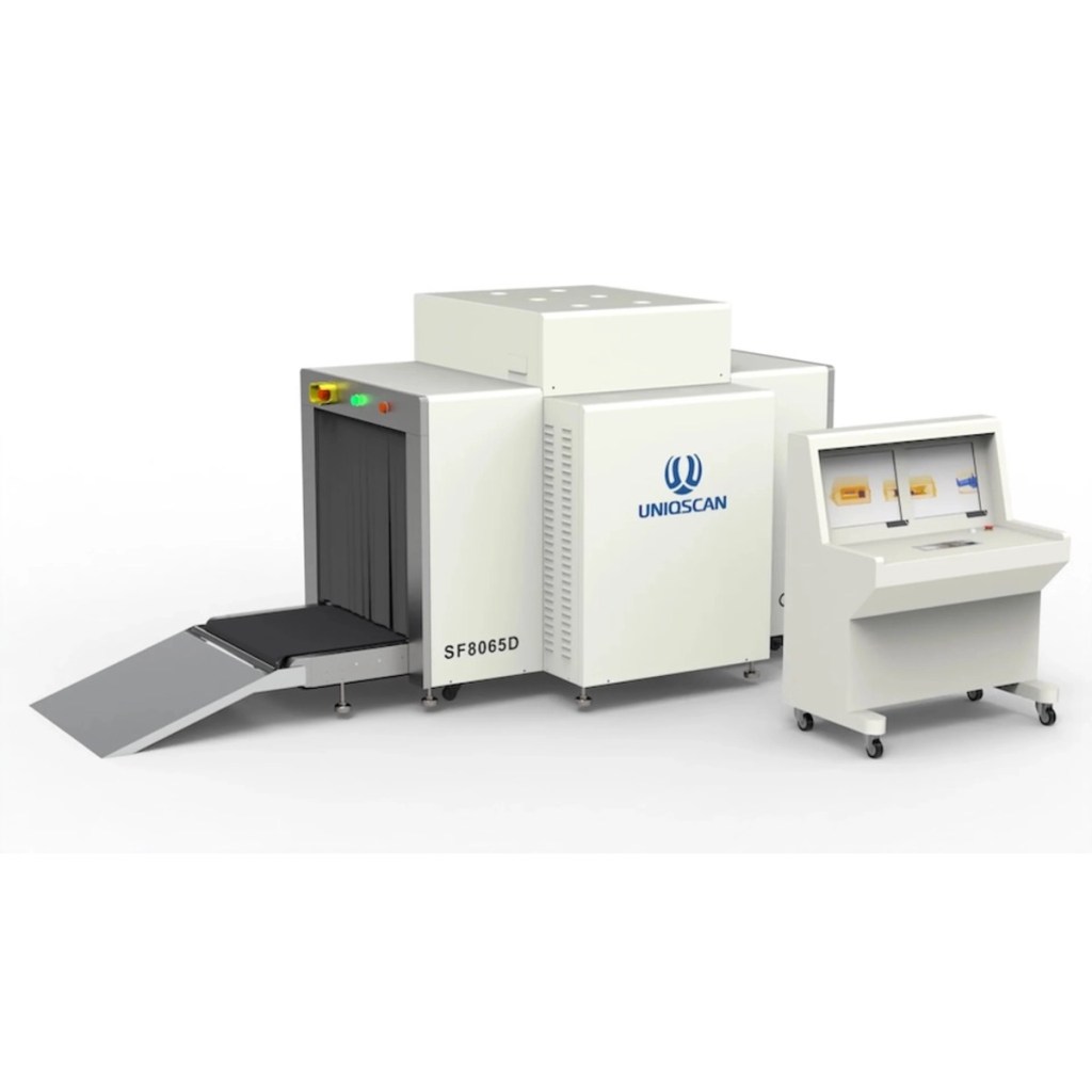

Modern advancements, like full-body scanners and colour-coded imaging systems, further enhance detection capabilities while maintaining passenger privacy. All these applications have significantly reduced the risk of threats and ensured the safety of travellers.
Applications in Industry and Beyond
Beyond medicine and security, X-rays have also found their application in other domains:
- Inspection & Quality Control: To inspect the integrity of materials, components, and welds in manufacturing, ensuring quality and safety.
- Mining: Analysing ore samples to determine the composition and purity of minerals. Precise data provided by X-rays aids in quick decision-making and profitability.
- Art and Archaeology: Used to examine artworks to verify their authenticity. In archaeology, X-rays are used to examine fossils and artefacts etc. without damaging them.
- Food Safety: Used to inspect food products for contaminants, metal objects etc. This ensures that they are safe for consumption.
Evolution
Over time the technology has evolved making it safer, quicker and more accessible. Early machines used cathode-ray tubes and photographic plates to capture images. These images were often of lower quality and required long exposure times.
Modern digital machines provide high-resolution images with shorter exposure times. This makes taking an X-ray safer than before with minimal radiation exposure. Traditional films have been replaced by digital film printers which can print the image in a matter of seconds. Digitisation also aids efficient storage and sharing of images.
Technology advances and miniaturisation have led to the development of Portable X-ray Devices. These devices are lightweight and easy to operate. This helps on-the-spot imaging in remote areas.
Inspiring Modern Medical Equipment
The discovery of X-rays laid the foundation for some of the most advanced medical imaging technologies in use today:
- CT Scan: Combines X-ray images taken from different angles to create detailed cross-sectional images of the body, providing more comprehensive information.
- MRI: X-rays inspired the development of MRI, another non-invasive imaging technique. MRI uses strong magnetic fields and radio waves to generate detailed images of soft tissues, organs, and structures within the body.
- PET Scan: PET scans use a combination of X-rays and radioactive tracers to visualize metabolic processes and detect abnormalities such as cancer.
Inspiring Modern Security Equipment
X-ray technology has also inspired innovations in security equipment:
- Full-body Scanners: Provide detailed images of individuals, revealing concealed objects and ensuring the safety of public spaces.
- Coloured Scanners: Uses colour coding to differentiate between types of materials, making it easier to identify prohibited items and potential threats.
- 3D Scanners: Creates 3D images of objects, providing more comprehensive information for security inspections.
These innovations have significantly improved security measures, enhancing the ability to detect and prevent threats.
The Future
The future of X-ray technology is as promising as its past. Emerging trends include:
- AI-Enhanced Imaging: AI algorithms are being used to refine image analysis leading to faster and more accurate diagnoses. This also reduces the risk of human error.
- Enhanced Imaging: This technique provides detailed insights into tissue composition. This will also provide greater detail and accuracy in images.
- Energy-Efficient Systems: New designs will reduce radiation exposure while maintaining image quality.
- Accessible Devices: Miniature devices will extend the coverage to farther and more remote areas.
- Personalised Medicine: Advanced imaging will analyse tissue composition at a molecular level. This will enable precise and personalised treatments based on individual disease data.
Legacy of Wilhelm Rontgen
Wilhelm Rontgen’s groundbreaking discovery earned him the first Nobel Prize in Physics in 1901. This highlights the transformative impact of his work on science and medicine. His legacy endures in the countless lives saved by this technology. It serves as a lasting inspiration for scientists and researchers, embodying curiosity, dedication, and innovation.
The story of X-rays is a testament to human ingenuity and determination. Starting with a chance discovery and evolving into a cornerstone of modern science. From diagnosing illnesses to enhancing security and revolutionising industries, they have profoundly reshaped the world. As we look ahead, their potential to drive innovations remains boundless, reflecting the enduring power of exploration and discovery.
I am sure you found this story interesting.
Please subscribe to my blog by filling in your details below:
My blog has countless such articles and stories to guide you and quench your thirst for knowledge.
You can also follow me on X and Facebook to read more such stories and posts.
PS: Copilot and ChatGPT have been used to create parts of this post.
Leave a Reply